CANDIDOSE (candidosis; synonymous with: candidamycosis, superficial blastomycosis, coormicosis, candidiasis, moniliasis, oidiomycosis) - an infectious disease of the skin, mucous membranes and internal organs caused by yeastlike fungi (fungi of the genus Candida).
The disease was first described by B. Langenbeck in 1839, although it has been known since the times of Hippocrates as stomata aphtoides and aphta infantis (K. Galek). In 1848, F. Th. Berg first identified the presence of yeastlike fungi in the tissues of a sick person. Berkhaut (M. Berkhaut) in 1923 among the yeastlike fungi distinguished genus Candida. The term Candida was adopted in 1939 by the III International Congress of Microbiologists.
Candidosis is found in all countries of the world, especially in the tropical and subtropical climate zone. The number of patients with various clinical forms of Candidosis, especially visceral, has some upward tendency.
Etiology
The causative agent of Candida albicans is most often Candida, less often C. tropicalis, C. krusei, C. pseudotropicalis, C. stellatoidea and some other species. The genus Candida (class Fungi imperfecti) includes over 80 species characterized by round, ovoid, less often cylindrical and sometimes irregular cells. Young cells - in diameter from 2 to 5 microns, mature cells - slightly larger. Yeastlike fungi do not have a true mycelium; they form chains (pseudomycelium) of elongated cells, which contact with each other with a narrow base (so-called strains), pseudomycelium length up to 12-16 microns. Cells are multiplied by germination and multipole bud (2-3 or more daughter buds). C. albicans and C. stellatoidea form spores at the ends of pseudomycelium with dense, usually double cell wall - chlamydopores.
Candidates are aerobes; they are often saprophytes of the mucous membranes of the mouth, intestines, vagina, and skin. They are found in soil, on fruits and vegetables. They use proteins, peptones and amino acids from nitrogenous substances for nutrition. Candidates are considered conditionally pathogenic microorganisms; their pathogenicity for humans and animals largely depends on the state of the macroorganism.
The most common for the cultivation of candidate (optimal temperature 30-37 °) are liquid medium Saburo, beer must, meat-peptone glucose agar. For the detection of chlamydospor most suitable medium Chapek - Doksa with the addition of twin-80, potato agar with bile, corn and rice agar.
Candidate retains its vitality in crops (dried out) for several years, tolerate repeated freezing and thawing in water and soil. They withstand competition with many microorganisms in terms of longevity on various products, such as sour milk, sauerkraut, fruit juices, etc. In cultures and pathogens, the material of fungi die by boiling within a few minutes. A 2-5% solution of phenol and formalin, chloramine, lysol, iodides, borates, copper and zinc sulphate, potassium permanganate and other chemical substances have a destructive effect; fungicidal effect is given by solutions of aniline dyes (gentianviolet, malachite green, methylene blue, etc.).
Epidemiology
The good adaptability of many species of the genus Candida to the environment ensures that they are widely distributed, as well as carried by humans and animals. For example, C. albicans can be found on the skin, mucous membranes and in the faeces of almost 20% of healthy people. Candidosis is found in calves, lambs, foals, poultry, etc.; diseases and wildlife are also observed. Some representatives of the genus Candida are associated with normal microflora of human skin (primarily C. albicans). As a source of infection, patients with fresh forms of skin and mucous membranes lesions are of the greatest importance. Exogenous infection occurs in direct contact with a patient or a fungal carrier (kiss, sexual contact, etc.) and through infected objects. Wastewater from baths, swimming pools and showers may, under unfavorable conditions, contribute to the disease of superficial foot candidiasis. Warm and humid climate, especially in summer, is important in the occurrence of candidiasis. Unfavorable working conditions (e.g., low level of technical equipment in confectioneries and fruit processing plants), violations of hygiene, both collective and personal, can even contribute to the occurrence of small outbreaks of superficial candidiasis in maternity hospitals, nurseries, confectioneries, etc.. There are known cases of infantile candidiasis of newborns during the passage of labor routes; very rare cases of congenital candidiasis are described. Endogenous infection is associated with the activation of yeastlike fungi already present in the microbial associations of the body, so in severe diseases (tuberculosis, pneumonia, diphtheria, malignant tumors, etc.) candidiasis may occur as a concomitant disease.
Pathological anatomy
Candidosis of the skin manifests itself as subacute dermatitis in the folds of the skin, rarely with the formation of abscesses and ulceration. In the histol, the study noted intercellular edema of the epidermis, paraceratosis, acanthosis and germination of threads of the fungus in the necrotized epidermis. Inflammatory infiltration mainly from segmental leukocytes is observed mainly in the dermis. Sometimes there is the formation of limited abscesses with the presence of decaying segmental leukocytes. Subsequently, granulomas are formed, consisting of lymphocytes, epithelioid and giant leukocytes.
In candidiasis of oral and pharyngeal mucous membranes, surface whitish-yellowish films are revealed. Microscopically, they consist of pseudo-mycelium fungus, slackened epithelium and a small number of segmental leukocytes. In more severe forms of lesions, the fungus penetrates between layers of the epithelium and parasites in the cells. There is dystrophy and swelling, perivascular inflammatory infiltrates in the derma.
Visceral candidiasis can be isolated (gastrointestinal tract, respiratory organs, genitourinary system) and generalized with single or multiple metastases in internal organs, nervous system, muscles, bones. Candidotic esophagitis is more often a consequence of the spread of the process from the mucous membrane of the mouth and yaw; three types of lesions are distinguished:
- Individual whitish plaque consisting of slackened epithelial cells, leukocytes and threads of the fungus, which are introduced between the cells of the multilayer flat epithelium;
- the formation of merging dense overlaps and the incorporation of the fungus into the submucosal layer;
- pseudomembranous overlaps that develop on the ulcerated mucous membrane; the threads of the fungus not only penetrate the necrotic masses, but also penetrate deep into the esophagus muscle and grow into vessels.
Affection of the stomach and intestines is less frequently observed, which is due to the peculiarities of the structure of the glandular epithelium, which produces sialomucines, and possibly the bactericidal properties of gastric juice. Usually, gastric and intestinal candidiasis develops against the background of previous inflammatory, ulcerative, atrophic changes. The sharper these changes are pronounced, the more severe the candidiasis takes. In the light form of filaments of the fungus only partially sprout epithelial cells and stele along the basal membrane, the inflammatory reaction is expressed poorly, mainly in the submucosal layer. In the severe form of mucous membrane necrosis is found, the threads of the fungus diffusely penetrate the necrotic masses and penetrate into the thickness of the stomach wall, intestines, this form may be accompanied by the formation of ulcers, which are sometimes complicated by bleeding or perforation with subsequent development of peritonitis.
Candidosis of the mucous membranes of the respiratory tract manifests itself as a catarrhal-desquamation and diphtheria inflammation. Candidotic pneumonia develops as a result of the spread of the process on the lung tissue along the length of the respiratory tract due to aspiration of the fungus or hematogenic way.
In the lungs, the early stage of inflammation caused by the candidate is characterized by exudate from segmental leukocytes with an admixture of fibrin. Further necrotic changes with passive fibrinous perspiration in alveolus predominate. In the chronic form of lung candidiasis produces multiple granulomas consisting of lymphocytes, histiocytes and giant cells, in which the cytoplasm often found phagocytic elements of pseudomycelium fungus.
Isolated urinary tract candidiasis is more likely to occur as a result of ascending infection; urethritis and cystitis are observed, which sometimes lead to pyelonephritis. In the purulent-necrotic foci of lesion, pseudomycelia fungus sprawl and a phagocytic reaction are detected.
Generalized candidiasis - single or multiple metastatic foci as a result of hematogenic spread of the fungus - may have different localizations. In case of brain lesion, pseudomycolium is widely spread around the vessels, which is associated with the peculiarities of the chemical composition of brain tissue, favorable for the fungus growth. Candidotic meningitis is characterized by acute limited or spilled purulent or productive inflammation.
Histol, the picture of changes in the metastatic foci of various organs and tissues has no strictly pronounced specific features. In the initial stage of the candidiasis process is observed necroticoexudative inflammatory reaction, more often purulent in nature, less often with a predominance of lymphocytes in the infiltration. The intensity of fungus growth depends on the viability of the tissue: where the necrosis is sharper, the threads of the fungus are much larger. In the case of chronus, the process around the necroticoexudative focus of inflammation involves the growth of connective tissue or the formation of granulomas similar to that of tuberculosis. Elimination of the fungus in the body is mainly due to lysosomal enzymes of segmental leukocytes, ie as a result of the so-called extracellular phagocytosis, so the growth of the fungus in the purulent focus is usually inhibited. The presence of a large number of segmental leukocytes prevents the fungus from multiplying, the leukocytes as if attaching elements of the fungus and, undergoing decay, isolate it from the surrounding tissue. As electron-microscopic studies show, in the cells of the fungus breaks the integrity of the cytoplasmic membrane and autophagous vacuum in the cytoplasm. Phagocytosis of fungi by segmented leukocytes is rare and affects mainly non-viable fungal cells; in the cytoplasm of macrophages and giant cells of the fungus can be a long time and be viable - the so-called endocytobiosis. Since the fungus is capable of intracellular parasitization, the presence of its viable forms indicates that the inflammatory process is prolonged.
In weakened, exhausted patients, inflammatory changes are very weak. This is especially noticeable if candidiasis is associated with diseases of the hematopoietic organs, in particular agranulocytosis and radiation disease.
Detection of candida on the surface of the skin, in the urethra, vagina and other mucous membranes should not always be considered a fungal disease. Candidosis is characterized by the presence of vegetative forms of fungus, the phenomena of kidney, filamentation, as well as centers of tissue necrobiosis with an inflammatory reaction and phagocytic fungus particles.
Pathogenesis
As a rule, candidiasis develops only when the body's defenses are weakened. Pathogenetic factors of this kind are manifold: hypofunction of the parathyroid and thyroid glands, disorders of carbohydrate metabolism, impoverishment of plasma with potassium at its normal content in red blood cells, dysproteinemia, violation of liver protein formation function, hypoadrenalism, dysmenorrhea, hypovitaminosis, maceration and minor skin injuries, chronic debilitating diseases.
Very important in the pathogenesis of candidiasis is dysbacteriosis, which develops through the use of antibiotics, corticosteroid hormones, cytostatics and immunosuppressants, which increases the fungal microflora and enhances its pathogenic properties. The immune state of the body and the degree of its specific sensitization, as well as the massiveness of the infection with fungi is essential.
The models of focal and widespread, acute and chronic forms of candidiasis of different localization have been developed on laboratory animals. Detection of fungi in small whitish nodules localized in the lungs, kidneys and spleen of the mice (rabbits on the 10th day) that died of candidiasis or were killed on the 5th day, is used to determine the degree of pathogenicity of the studied cultures. Extracts from the cells of freshly isolated fungi, which were the most pathogenic for animals, have expressed immunogenic properties. Polysaccharide preparations obtained from the treatment of yeast cells with pepsin, betanafluoroids and ethanols were most suitable for serum, candidiasis diagnostics, immunodiffusion reaction in gel, complement binding reaction, indirect hemagglutination and latex particle agglutination reaction. In the course of infection and immunization, live and killed vaccines produce agglutinins, precypitins, antibodies that bind the complement. Phagocytosis is also intensified, and allergic restructuring of the body occurs.
Immunity
There is no congenital immunity to candidiasis; acquired immunity is weak. Healthy people very rarely get candidiasis, especially visceral form, although the candidates have allergic properties, and their entry into the body occurs in infancy. In healthy people, the titer of agglutinins ranges from 1:20 to 1:160.
Classification
Due to the great variety of manifestations of candidiasis many classifications are proposed. However, by clinical manifestations of candidiasis can be divided into four main groups.
A. Superficial Candidosis:
- Candidosis of mucous membranes: a) yeast stomatitis (thrush); b) yeast glossitis; c) candidiasis angina; d) candidiasis vulvovaginitis; e) candidiasis balanitis and balanopositis.
- Candidosis of skin: a) candidiasis; b) candidiasis heilitis; c) Candidosis of large folds of skin (Interrigenous Candidosis, Interrigenous Yeast Mycosis); d) Interfinger candidiasis of brush erosion; e) Candidosis of small folds of skin; f) other manifestations of smooth skin. 3. Candidosis of nail rollers and nails.
B. Chronic generalized (granulomatous) Candidosis.
C. Visceral Candidosis:
- Candidosis of respiratory tract.
- Candidosis of digestive tract organs.
- Candidosis of urinary system.
D. Secondary (allergic) forms of candidiasis.


Clinical symptomatology
Candidosis of mucous membranes
Yeast stomatitis, or so-called thrush stomatitis, is found mainly in weakened infants and young children, or in elderly people against the background of chronic diseases. Initially, on the invariable mucous membrane of the cheeks, hard and soft sky, tongue and gums are formed dotty deposits of white color, which, merging, form white films, reminiscent of collapsed milk. If you remove the films, underneath them there is a bleeding mucous membrane. In case of thrush, a child may have a nipple candidiasis in a breastfeeding woman: the skin turns red, there are bubbles and cracks on the nipple, sharp pain in feeding.
Yeast glossitis - in addition to the thrush, in some areas of the back of the tongue can be observed atrophy of the filamentous nipples, some patients are significantly pronounced deep grooves of the tongue, in which you can see the whitish color of plaque, the whole tongue is slightly increased due to swelling. Candidosis of the mucous membranes is not a serious disease, but it is a symptom indicating a general weakening of the body; yeast glossitis is often observed in elderly people wearing dentures.
Candida sore throat is usually chronic with normal body temperature and no soreness when swallowing. Whitish plugs are formed on amygdala, but there may also be plaque in the form of white films. Regional lymph, nodes are not involved in the process.
Candida vulvovaginitis is detected by the appearance of whitish secretions, which have a tiny character. On the mucous membrane of the vagina, which is usually hyperemic, there are whitish or gray plaque, surface erosions. Patients note itching, burning in the area of the external genitalia. Often, candidiasis is combined with trichomonadic colpitis.
Candidotic balanitis and balanopostitis - on the head of the penis, on the inner sheet of foreskin and less often on the outer sheet are white patches, under which surface erosions are found, rashes are accompanied by burning, painfulness.
Candidosis of skin
Candidate seizure may occur as an independent disease or be a manifestation, for example, thrush. Yeast infection is characterized by predominance of maceration and delamination of the upper corneal layer in the form of a kerb around the cracks in the corners of the mouth and significant infiltration at the base.
Candidate heilite is characterized by red red edging of the lips, dryness, a sense of tightening and burning. The scales are grayish, as if glued to the red lip border, and their free edge slightly rises; to diagnose candidiasis heilitis, it is necessary to re-discover the fungus elements.
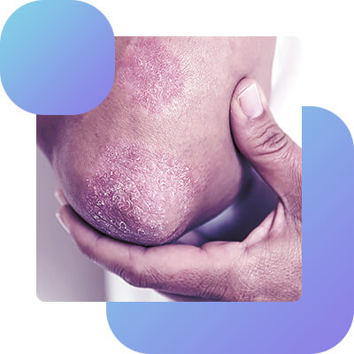
Intrigenous candidiasis (large folds) is quite common. It may affect the armpit pits, skin folds under the mammary glands, especially in obese women, buttock and inguinal hip folds. This localization creates large hearths with clear boundaries, often with erosions and wetness. The periphery of the hearths is surrounded by a curb of whitish macerated epidermis. Around the main hearth, especially under the breast, are often found small hearths of the same nature (the so-called daughter elements).
Interfinger candidiasis erosion of the hands is very common, sometimes as a manifestation of occupational disease (professional candidiasis). Usually, the process develops in the third interfinger fold, less often in others. The corneal layer in the fold swollen, macerated, has a pearlescent shade. In the center of the hearth is found an eroded surface of red, wet and shiny due to moderate wetness. Further on the side surfaces of the main phalanges the process does not extend. Erosions occur quite persistently and without elimination of pathogenetic factors are prone to relapse. Patients note itching and burning.
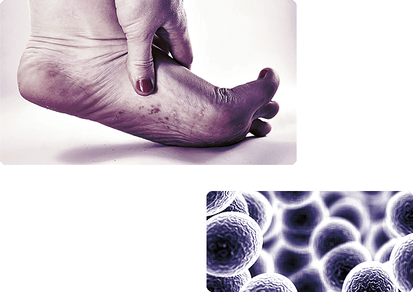
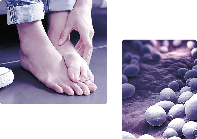
Candidosis of small skin folds (behind the auricles, in the navel area, interfinger folds of the feet) is common. The process is characterized by the same wedges, signs that are localized in large folds, and may be either the primary manifestation of candidiasis or develop along with the signs of another disease.
Other manifestations of smooth skin candidiasis may be found in the form of erythematosquamosis, vesiculobullosis, scarlet-like and psoriasiform rashes. Candida erythroderma is also rarely observed.

Candidosis of nail rolls and nails (onychia and paronichia) is usually observed only in the hands, as a professional disease in the workers of fruit processing plants.
The process begins more often with a nail roll, which becomes hyperemic, "pillow-shaped", disappears the cuticle, when pressed, a small drop of pus is released, an acute stage gradually becomes subacute and chronic. Later, the nail plate is involved, which gradually becomes bumpy with transverse stripes and pressure, or thin, sometimes easily peeling off. The color of the affected nail records is brownish, less often with a greenish hue. Yeast paroniasis and onychia may be the only manifestation of candidiasis or combine with other forms of surface or even visceral candidiasis.
Visceral Candidosis
Chronic generalized (granulomatous) candidiasis is an independent wedge, a type of candidiasis. As a rule, the disease begins in early childhood with the thrush of the oral mucosa. Then, gradually the nail rolls and nail plates of the hands and feet, pilose scalp, face, body and limbs are involved. Frequently observed yeast heilitis, especially lesions of the lower lip, leads to the development of macroheil. Hyperemic and peeling spots with infiltration of the base, papules, tubercles and horn deposits are characteristic of the skin of the scalp, face, torso and extremities. Many patients have recurrent pneumonia: and epileptiform seizures, possibly lesions of the liver and kidneys. The course of the disease is chronic with exacerbations. Most of the patients are exhausted, children lag behind in physical development.
Candida panophthalmia, irrite, endocarditis, etc. are observed.
Candidosis of the respiratory tract may be primary or secondary. Affection of the throat and larynx is accompanied by dry cough attacks, change in the tone of voice, laryngostenosis and sputum secretion. Candidotic bronchitis is manifested by persistent coughing, mucosa purulent sputum, large bubble wheezing. Primary yeast pneumonia more often occurs in the treatment of any other disease with antibiotics, and the secondary may occur in patients with tuberculosis and proceed as a complication of pneumonia of another etiology. Sometimes, due to the formation of caverns, mycotic pneumonia resembles tuberculosis lesions of the lungs, which should always be considered when making a differential diagnosis. Sometimes it resembles lung sarcoidosis.
Candidosis of the digestive tract is manifested by various symptoms: a decrease in appetite, difficulty swallowing, vomiting with the release of cottage cheese films, liquid stool with an admixture of mucus, etc. With the progression of bowel candidiasis dehydration occurs, expressed intoxication, adynamy, fever may be observed.
Candidosis of the urinary system is accompanied by the appearance of protein, blood and cylinders in the urine, an abundance of fungus elements; renal filtration capacity is impaired.
Secondary forms of candidiasis
Secondary forms of candidiasis (levurids, or mikides) occur when there is a primary focus in the internal organs, skin or mucous membranes, which sensitize the body. Clinically, levurids occur in the form of erythematosquamosis, vesicular, parapsoriaziform or other elements. Secondary allergic rashes are usually symmetrical and may be accompanied by headache, sickness, heart disorder, and a change in the formula of peripheral blood. In the occurrence of levurids are essential disorders of the treatment regimen, irrational therapy, trauma to the centers of candidiasis or chemical irritation.
Complications vary, but the most severe are septicopia and septicemia.
Diagnostics
The diagnosis of candidiasis of visible mucous membranes, skin, nail rolls and nail plates is made on the basis of wedges, manifestations and presence of fungi in the examination of the material from the surface of the focus areas. In visceral candidiasis, additional examinations are required, as well as contamination of laboratory animals.
Laboratory tests. Material for examination are skin and nail scales, detachable ulcers, pus, cerebrospinal fluid, blood, urine, bile, faeces, pieces of biopsy tissue and cadaver material. Pathol, material is microscopic in 10% solution of caustic alkali, or in solution of Lugol double fortress, or in a mixture of alcohol and glycerol (2 tsp. alcohol, 4 tsp. glycerol, 4 tsp. water).
Candidate can be found in histol. research - coloring slices of lesions by Gram Weigert in various modifications. They are best detected when colored on neutral mukopolysaccharides by Chochkiss-Mac-Manus or by Shabadash: a uniform coloring of yeast-like cells and pseudomycelium of the fungus is observed, phagocytic elements of the fungus and dying particles of the fungus are well detected. In the early stages of parasitization, the direct method of luminescence (fluorescent antibodies) is used to detect the candidate. As the inflammatory process develops, the intensity of fungus luminescence decreases, which is associated with the synthesis of antibodies against fungus antigens. In tissue slices, the fungus is found in the form of round or oval yeastlike cells, sometimes budding, pear-shaped. Side budding is often seen at the junction of cells (turbidity). As a result of filamentation (thread formation), thin, short, curved and long filaments of pseudomycelium are observed, sometimes with thickening at the ends to 7 microns thick.
Cultural diagnostics is achieved by sowing pathols, material on the medium Saburo and wort with streptomycin, penicillin, left micacetin to inhibit the growth of accompanying microbes. For the detection of pseudomycelium and the formation of chlamydospor, the most suitable are dashed 4-5-day crops on beveled carrot, corn or rice agar environments. The growth of a large number of yeast colonies (more than a thousand per gram of the studied material in crops on dense media) suggests that the organism may be ethyl, an agent of the disease; however, repeated sowing of the same studied material is necessary.
Serol, diagnosis of candidiasis is achieved by the agglutination reaction and the complement binding reaction. A strongly positive agglutination reaction in serum dilution of at least 1 : 200 is considered reliable, and the reaction gives clearer results with the autoantigen, a culture obtained from a patient. The reaction of complement binding is more specific; the clearest results are obtained with generalized and visceral forms.
Specific immunofluorescence has also proved useful for serotype identification and candidate identification.
The most important for the diagnosis of candidiasis is the increase of antibody titers in the course of the disease; the increase in the number of yeastlike fungi in repeated studies of pathogens, material. Positive allergic skin reactions are important in combination with other data.
Differential diagnostics between candidate, aspergillus and nocardias should be performed.
Treatment
The therapy is carried out with mandatory consideration of pathogenetic factors. For example, the normalization of carbohydrate metabolism or thyroid function, improvement of the general condition of the body may lead to the elimination of clinical manifestations of candidiasis. Means of etiol, effects accelerate recovery. General tonic and proper nutrition of patients are important: the food should be rich in proteins, vitamins, but the amount of carbohydrates should be limited. It is shown the prescription of repeated courses of nystatin, levorin or their salts (both antibiotics are most effective in case of candidiasis of the digestive tract). Nystatin is administered orally to adults 6 000 000 - 8 000 000 units per day (between courses there are breaks). Amphotericin B is shown in case of severe visceral candidiasis and generalized chronic candidiasis; it is prescribed intravenously in 5% glucose solution in the form of drip injections at the rate of 250 units per 1 kg of body weight for adults with a total course dose of 1 500 000 - 2 000 000 000 units. Immunotherapy for patients with candidiasis is widely used; a polyvalent vaccine (or outovaccine) is prescribed. Fungus C. albicans containing a complex of proteins, polysaccharides and lipids are also used.
External therapies are varied. 1-2% alcohol or aqueous solutions of aniline dyes, Castellani liquid, benucid, mycoseptin, zinccondane and other fungicidal liquids are successfully used. Amphotericin, levoricin and nystatin, 10% sulfuric and 3% salicylic are used in ointments. In case of candidiasis of the oral mucous membrane, it is recommended to apply smears (2-3 times a day) with aqueous solutions of aniline dyes, 10-20% borax solution in glycerine, rinsing with 1-2% aqueous solution of tannin or 1% aqueous solution of iodinol, and tablets with decamel as caramel.
To treat children, use 1-2% solution of (aqueous) aniline dyes, 5-10% solution of glycerine borax - greasing the hearths 3-4 times a day; mouth rinsing 5-10% solution of tannin, and caramel and deacon.
At vulvovaginitis use tampons with 20% solution of borax in glycerine, foaming tablets of levorine, lubrication of 1-2% with water solution of aniline dye.
Forecast
With surface forms, the prognosis is favorable; for example, breast cancer in infants with proper treatment and nutrition is cured quickly. The prognosis is more serious in chronic generalized and visceral form, especially in allergic complications. To a large extent, the prognosis depends on the course of the disease, against which candidiasis has developed.
Prophylaxis
In the production of fruit and vegetables, juices and syrups processing it is necessary to implement mechanization and automation that excludes the use of manual processing. In case of infectious and other diseases, long-term use of antibacterial antibiotics should be combined with nystatin (up to 400 000 - 600 000 units per day) and a complex of vitamins, especially vitamin B. It is recommended to use rubber slippers in baths and showers. Especially important is the prevention of newborns and infants; in maternity hospitals and nurseries, people with signs of candidiasis should not be allowed to work with children. In the home, people with candidiasis should avoid communicating with children; dishes, linen, and towels should be disinfected.
By: Dr. Susan Bard